Diagram Of Shoulder Bones - Shoulder Bones - Joint Preservation Center - Neck vertebrae (7) (cervical vertebrae).. 9 photos of the shoulder bones anatomy diagram. The first type is the white cartilage on the ends of the bones (called articular cartilage) which allows the bones to glide and move on each other. Click now and learn everything about its anatomy and function at kenhub! Shoulder problems including pain, are one of the more common reasons for physician visits for musculoskeletal symptoms. Placebo injection for adhesive capsulitis of shoulder.
Her gün binlerce yeni, yüksek kaliteli fotoğraf ekleniyor. Shoulder problems including pain, are one of the. Shoulder bones and ligaments anatomy. An example of shoulder flexion can be seen when reaching forward to grasp an object. However, it is an unstable joint because of the range of motion allowed.
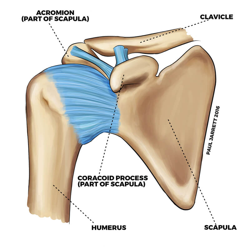
Shoulder Anatomy | Dr Paul Jarrett, Hand, Wrist & Shoulder ... from pauljarrett.info Human science human body arm anchor chart bone shoulder health skeleton diagram. The bones of the shoulder consist of the humerus (the upper arm bone), the scapula (the shoulder blade), and the clavicle (the collar bone). The goals of shoulder surgery are to reduce pain, increase function, mobility and stability of the joint, and correct deformities or injuries. Several muscles that originate at the posterior surface of the ulna or the radius (the other bone in the forearm) have their actions in the hand. Cheek bone (zygoma) upper jaw (maxilla). Scapula (= 'shoulder blade' or 'shoulder bone') is a bone of the human body. Shoulder bones and ligaments anatomy. Shoulder problems including pain, are one of the.
Lower jaw (mandible) collar bone.
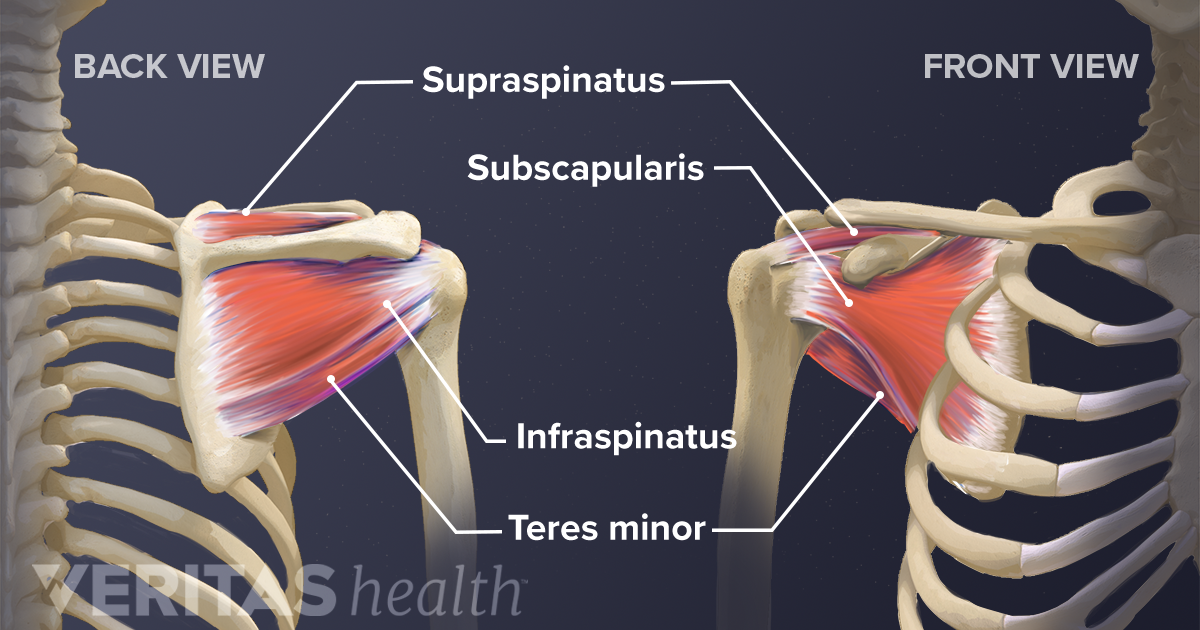
The shoulder joint (glenohumeral joint) is a ball and socket joint between the scapula and the humerus. The rotator cuff muscles are four muscles that form a musculotendinous unit around the shoulder joint. Neck vertebrae (7) (cervical vertebrae). Shoulder flexion is movement of the shoulder in a forward motion. The shoulder is not a single joint, but a complex arrangement of bones, ligaments, muscles, and tendons that is better called the shoulder girdle. Due to this, there is a hollow centre inside the backbone. 9 photos of the shoulder bones anatomy diagram. The shoulder bones, rib bones and hip bones ,are all joined to the backbone. Lower jaw (mandible) collar bone. Most relevant best selling latest uploads. It is made up of three bones: When this type of cartilage starts. This image shows the shoulder joint displaying the bones and ligaments that form the joint and supports it (from anterior view) showing:
Each vertebra has a hole in it. The shoulder bones, rib bones and hip bones ,are all joined to the backbone. Labeled human shoulder bone anatomical vector illustration diagram poster. Shoulder flexion is movement of the shoulder in a forward motion. .diagram shoulder blade bone shoulder bones and ligaments right shoulder joint anatomy back shoulder bones anatomy of shoulder rotator cuff human scapula anatomy left shoulder bones shoulder joint tendons upper arm bone anatomy frozen shoulder anatomy shoulder.
Soft Tissues of the Shoulder from embed.widencdn.net Her gün binlerce yeni, yüksek kaliteli fotoğraf ekleniyor. However, it is an unstable joint because of the range of motion allowed. Shoulder flexion is movement of the shoulder in a forward motion. Each vertebra has a hole in it. The transverse humeral ligament is not shown on this diagram. Shoulder joint is the most mobile joint of the human body. The main functions of shoulder bone are : The goals of shoulder surgery are to reduce pain, increase function, mobility and stability of the joint, and correct deformities or injuries.
Shoulder diagram illustrations & vectors.
This page is about shoulder bone anatomy diagram,contains anatomy of the shoulder central coast orthopedic medical group,anatomy of the shoulder part 3 (muscular structures),shoulder replacement,guide to shoulder anatomy and more. The shoulder is the most movable joint in the body. Posterior to the clavicle is the scapula, a flat, triangular bone located lateral to the thoracic spine in the dorsal region of the body. Labeled human shoulder bone anatomical vector illustration diagram poster. The rotator cuff muscles are four muscles that form a musculotendinous unit around the shoulder joint. Several muscles that originate at the posterior surface of the ulna or the radius (the other bone in the forearm) have their actions in the hand. Download 708 shoulder diagram stock illustrations, vectors & clipart for free or amazingly low rates! The articulations between the bones of the shoulder make up the shoulder joints. The scapula is a large, flat triangular bone with three processes called the acromion, spine and coracoid process. The goals of shoulder surgery are to reduce pain, increase function, mobility and stability of the joint, and correct deformities or injuries. Shutterstock koleksiyonunda hd kalitesinde diagram showing shoulder bones illustration temalı stok görseller ve milyonlarca başka telifsiz stok fotoğraf, illüstrasyon ve vektör bulabilirsiniz. (1) collar bone on the two sides of the next keep our shoulders apart. There are two kinds of cartilage in the joint.
A normal, complete, human skeleton includes two shoulder blades, which the scapula is a flat bone so it is conveniently described by two diagrams, one to label the features of the posterior surface of the scapula and the. The shoulder joint (glenohumeral joint) is a ball and socket joint between the scapula and the humerus. Following inferior dislocation of shoulder joint, the rounded contour of shoulder is lost and there is weakness of abduction of armbecause the. Shoulder problems including pain, are one of the. The transverse humeral ligament is not shown on this diagram.
Anatomy and Function of the Shoulder | Smith & Nephew - US ... from www.smith-nephew.com This image shows the shoulder joint displaying the bones and ligaments that form the joint and supports it (from anterior view) showing: The articulations between the bones of the shoulder make up the shoulder joints. Due to this, there is a hollow centre inside the backbone. This page is about shoulder bone anatomy diagram,contains anatomy of the shoulder central coast orthopedic medical group,anatomy of the shoulder part 3 (muscular structures),shoulder replacement,guide to shoulder anatomy and more. These new cartilage cells push older, larger cartilage cells towards the middle of a bone. Shoulder diagram illustrations & vectors. .diagram shoulder blade bone shoulder bones and ligaments right shoulder joint anatomy back shoulder bones anatomy of shoulder rotator cuff human scapula anatomy left shoulder bones shoulder joint tendons upper arm bone anatomy frozen shoulder anatomy shoulder. In human anatomy, the shoulder comprises the part of the body where the arm attaches to the torso.
These new cartilage cells push older, larger cartilage cells towards the middle of a bone.
Shoulder flexion is movement of the shoulder in a forward motion. Several muscles that originate at the posterior surface of the ulna or the radius (the other bone in the forearm) have their actions in the hand. However, it is an unstable joint because of the range of motion allowed. Her gün binlerce yeni, yüksek kaliteli fotoğraf ekleniyor. In human anatomy, the shoulder comprises the part of the body where the arm attaches to the torso. Shoulder bones and ligaments anatomy. Download 708 shoulder diagram stock illustrations, vectors & clipart for free or amazingly low rates! The shoulder is the most movable joint in the body. The main functions of shoulder bone are : The articulations between the bones of the shoulder make up the shoulder joints. When this type of cartilage starts. The scapula is the flat triangular bone which provides an attachment for muscles for the shoulder, back and neck. Placebo injection for adhesive capsulitis of shoulder.
It is made up of three bones: diagram of shoulder. Neck vertebrae (7) (cervical vertebrae).


0 Komentar